Definition. A localized defect in the visual field bordered by an area of normal vision. This occurs with a variety of EYE DISEASES (e.g., RETINAL DISEASES and GLAUCOMA), OPTIC NERVE DISEASES, and other conditions. [

Keeping this in view, what is an arcuate visual field defect?

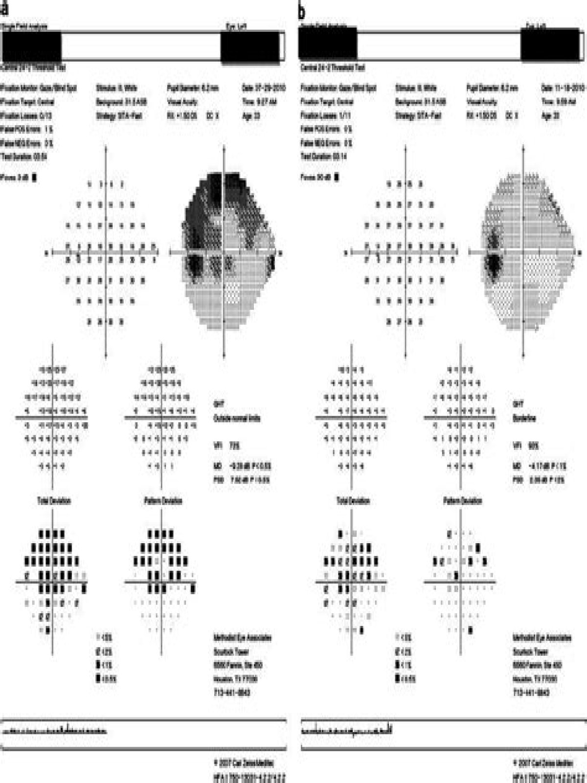
Glaucomatous damage to a nerve fiber bundle containing axons from both the inferonasal and inferotemporal retina resulted in the arcuate defect shown. The scotoma often begins as a single area of relative loss, which then becomes larger, deeper, and multifocal.

Similarly, what does it mean if you have large optic nerves? The cupping of the optic nerve means the size of the depression in the middle of the nerve when viewed from the front of the eye. When a person is shown to have large optic nerve cups, it could be an indicator of damage unless it can be determined that the cup size is considered normal for that individual.

In this way, what causes a scotoma?

A scotoma is caused by a problem in your brain, a problem in your eye, or a problem in your optic nerve. The optic nerve is located behind your eye and sends pictures to the brain. The kinds of problems that can cause a scotoma include: A stroke.

Can scotoma be cured?

Unfortunately, scotomas cannot be corrected by glasses or contact lenses. Sometimes surgery may be helpful in treatment of a scotoma. For example, if the scotoma is caused by a tumor, removal of the tumor may correct the scotoma.

How fast does glaucoma progress?

Untreated acute glaucoma results in permanent vision loss. Untreated chronic glaucoma can progress to blindness within several years. Early diagnosis and treatment usually results in excellent success and vision is saved.

What can be mistaken for glaucoma?

Conditions that can be mistaken for glaucoma include compressive or infiltrative lesions of the optic nerve, previous ischemic optic neuropathy (both arteritic and non-arteritic), congenital and hereditary optic neuropathies, post-traumatic optic neuropathy and inflammatory and demyelinating optic neuritis.

Can an optometrist detect glaucoma?

All optometrists, no matter their level of licensure, are expected to diagnose the condition or at least to recognize that a problem exists and refer the patient to another practitioner. It is in the area of the diagnosis that most referrals by optometrists to ophthalmologists related to glaucoma occur.

What makes you a glaucoma suspect?

Glaucoma suspect describes a person with one or more risk factors that may lead to glaucoma, including increasing IOP, but this person does not yet have definite optic nerve damage or vision loss due to glaucoma. By monitoring them for the earliest signs of glaucomatous damage, visual function can often be preserved.

What is the first sign of glaucoma?

If the entire optic nerve is destroyed, blindness results. Other symptoms usually are related to sudden increases in IOP, particularly with acute angle-closure glaucoma, and may include blurred vision, halos around lights, severe eye pain, headache, abdominal pain, nausea, and vomiting.

Can you have glaucoma in just one eye?

Primary open angle glaucoma generally affects both eyes, but does not always develop at the same time. One eye may have moderate or advanced glaucomatous damage, while the other eye has very little or none. Some types of glaucoma such as secondary glaucoma caused by injury to the eye, usually affect one eye.

Can anemia cause glaucoma?

Iron deficiency anemia is an important public health problem. Also it is considered to be a risk factor for many diseases. The study demonstrates the iron deficiency anemia frequency in glaucoma patients and compares with the normal subjects.

What is Bjerrum scotoma?

Bjerrum's area is the central 25° of the visual field from the fixation point, popularized scientifically by the Danish ophthalmologist Jannik Petersen Bjerrum. Of particular note is the arcuate scotoma (also known as the Bjerrums scotoma).

Are Scotomas dangerous?

It may be difficult to read and dangerous to drive a vehicle while the scotoma is present. Normal central vision may return several minutes before the scotoma disappears from peripheral vision.

Do Scotomas get worse?

Patients present with rapid onset of central vision loss that may be described as blurred vision, paracentral scotoma, metamorphopsia, “spots” in the vision, and photopsias. Initial vision at presentation is 20/25 or worse in about 77% of eyes and 20/40 or worse in 58%.

How long does scotoma last?

The visual symptoms typically last approximately 20-30 minutes and then completely resolve. The area where vision is disrupted is known as a 'scotoma' and the whole episode is often referred to as an 'aura. '

How is scotoma diagnosis?

The presence of the blind spot scotoma can be demonstrated subjectively by covering one eye, carefully holding fixation with the open eye, and placing an object (such as one's thumb) in the lateral and horizontal visual field, about 15 degrees from fixation (see the blind spot article).

Do Scotomas move?

A scotoma is a blind spot in your vision. It can be temporary or permanent, and it may stay in the same place or move around in your vision.

What does the blind spot look like?

Blind spot, small portion of the visual field of each eye that corresponds to the position of the optic disk (also known as the optic nerve head) within the retina. With both eyes open, the blind spots are not perceived because the visual fields of the two eyes overlap.

Can stress cause an aura?

The cause of migraine with aura isn't entirely understood. Many of the same factors that trigger migraine can also trigger migraine with aura, including stress, bright lights, some foods and medications, too much or too little sleep, and menstruation.

How do u know if ur going blind?

10 signs and symptoms of eye problems
  • A sudden onset of many spots and floaters in your field of vision.
  • A sensation that a dark curtain has settled across your field of view.
  • Sudden eye pain, redness, nausea and vomiting.
  • Sudden blind spot in one eye.
  • Poor night vision, halos around lights or less vivid color vision.

Where is the blind spot located?

The blind spot is the location on the retina known as the optic disk where the optic nerve fiber exit the back of the eye.